Massachusetts Eye and Ear Using Smartphones For Medical Photos

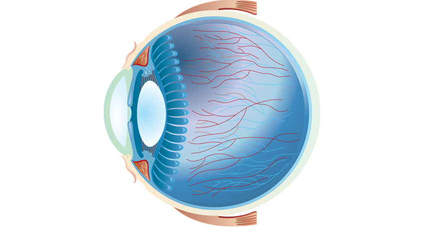
Eye image via shutterstock
In a paper published in the Journal of Opthalmology last week, ophthalmologists at Massachusetts Eye and Ear Infirmary discussed the benefits of using iPhone cameras, with the help of medical lenses and an enhancing app, to take medical eye photos.
Fundus photography, the photo that the ophthamologist takes of the interior surface of your eye, is a key component in ophthalmology. Doctors at Mass. Eye and Ear explained in their paper that the simple combination of an iPhone, an app, and medical instruments found in ophthalmologists’ offices are transforming medical practices.
Dr. Shizuo Mukai, a senior author of the article and an associate professor at Harvard Medical School said in a press release:
“This technique has been extremely helpful for us in the emergency department setting, in-patient consultations, and during examinations under anesthesia as it provides a cheaper and portable option for high-quality fundus-image acquisition for documentation and consultation.”
On both children and anesthetized adults, researchers used either an iPhone 4 or 5, a $4.99 app called Filmic pro, and a 20D lens or a Koeppe lens — a special curved lens that allows the doctor to see the back of the eye — to produce high quality images of the patients’ eyes.
Filmic pro enhances the iPhone quality images and allows doctors to adjust the focus, exposure, and light intensity while taking photos and videos. This gives the doctors plenty of control over the image and makes fundus photography portable.
The paper says researchers achieved the best results in an operation room using a Koeppe lens in addition to a 20D lens, but photos with just a 20D lens in the emergency room and waiting rooms were excellent as well.
This technique is cheap and simple, and takes advantage of the prevalence of smartphones for medical technology advancement, the paper says.


