New Non-Invasive Skin Biopsy Technology Makes Doctor Visits Easier

Image provided
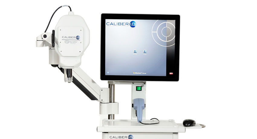
Medical device company Caliber I.D. has developed a new gadget that will change the way you think about going to the doctor. The VivaScope system is a high-resolution medical imaging system that allows doctors to perform skin biopsies, to test for cancerous cells in moles, for example, without cutting into a patient’s skin.
Caliber, which used to be called Lucid Technologies, started to develop a laser hair removal system in the early nineties that used a laser imaging system to get down to cellular structure of each individual hair follicle. However, so many similar systems were being developed at the time, that funding was limited. Federal funding was available, however, for skin biopsy laser technology. So Lucid jumped switched tacks. Teaming up with Massachusetts General Hospital researchers, Lucid used the same kind of technology to produce the first VivaScope system in 1997.
“Basically its a low-powered laser,” explains Michael Hone, CEO of Caliber I.D. “It doesn’t have much more power than a laser pointer. We shine the laser on the skin and it uses the luminescence of the skin to reflect back a picture at the cellular level.”
Hone says the technology is akin to shining a flashlight under your hand and seeing it reflect through your skin. “Except here we’re pinpointing the light with much more accuracy in order to get down to the individual cell,” he says.
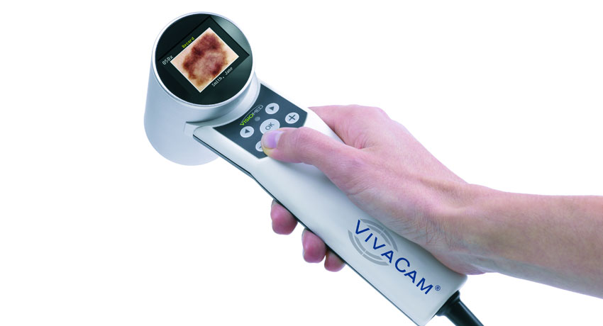
With a quick point and shoot, the VivaScope magnifies the selected area of skin and projects an image of the junction between the epidermis and the dermis. This image is displayed on a touch screen, not unlike the one on your iPhone, and doctors can examine the selected area of skin there. Trained dermatologists and pathologists can very easily tell if there is something wrong with the biopsied skin just by looking at this image. Hone says VivaScope is making it possible to make bedside diagnoses, something that was previously unheard of in skin biopsies.

Image provided
For a traditional skin biopsy, a dermatologist will numb the specified area, excise a piece of skin, and send it off to a pathology lab. The pathologists in the lab then freeze, section, and stain the skin sample and look at it under a microscope. Results could take up to a week. VivaScope eliminates most of this procedure, allowing real time diagnoses.
“Obviously our system is a lot simpler,” Hone says.
If there is a question about the cells in the specified area, dermatologists can share the images they collect from VivaScope over an interconnected database called VivaNet. While the patient is still in the office, dermatologists can share the skin image with pathologists in separate labs who can help to make a more specified diagnosis.
Once a VivaScope “optical biopsy” is completed and the sample is determined to be concerning, dermatologists can use the image that VivaScope provides to ensure clean margins when they cut out the worrisome tissue. This helps to make sure that not cancerous cells come back later on.
It all sounds great, but is it more expensive for the patient? Hone says that for the time being, it is. “It’s about the same cost as a conventional biopsy,” he says, “but its an out of pocket expense at the moment. We’re working with insurance companies now and we hope to have it covered under most insurance plans in the next year.”
Hospitals like Massachusetts General Hospital, who helped develop the original technology in the 90s, Brigham and Women’s, Johns Hopkins, and the Mayo Clinic are using VivaScope technology regularly.
Hone says European doctors have adopted the technology quicker. “Especially Germany,” he says. “Germany has already included VivaScope in their standard of care guidelines so it is available to everyone. In their case, its covered by insurance as well, like we hope to have here in the coming months.


