Somewhere Over the Brainbow

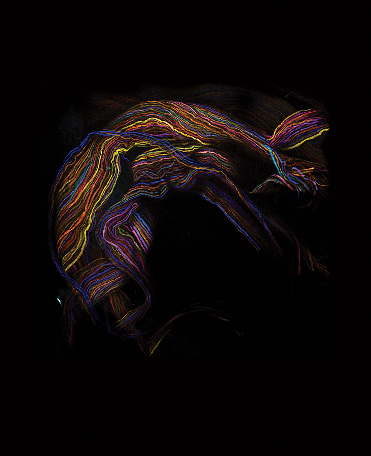
image courtesy of Livet & Lichtman
This image traces the pathways of nerve impulses in a mouse’s body. Scientist Jeff Lichtman used genetically engineered mice and fluorescent proteins to turn maps of their brains’ nerve fibers into psychedelic blacklight posters. For the first time, scientists looking at slivers of mouse brain can tell where individual nerve fibers, called axons, go and how they connect the mouse’s neurons. Lichtman calls this technique the “Brainbow” method—for obvious reasons.
“So there are these deep mysteries of outer space, and there’s an even deeper mystery very close by in every single person—on top of their shoulders. We understand the liver, the kidneys, the spleen, the lungs, the heart, the digestive system, the skin. But when you come to the brain, you’re confronting a physical tissue that is just vastly more complicated than anything else.
The machine you’re trying to understand is more elegant, subtle, and complicated than the rather crude ideas that come out of our brains, no matter how nuanced they are. And this is a big crisis for biologists: We’re trying to understand something that’s more complicated than the way we think.
When we think of scientists, we think of people who are generating objective data; they want to turn everything into numbers and graphs and do statistical analyses. But in the pathway toward analysis you have to generate a rendering of the data, and the rendering we use is a visual, and the rendering turns out to be—oh my God, just beautiful!
Humans have some kind of appreciation for natural beauty. But why should the inside of the brain, which is something we have never come in contact with before, be so awesome visually and stimulating? That’s a deep question.” —As told to Janelle Nanos

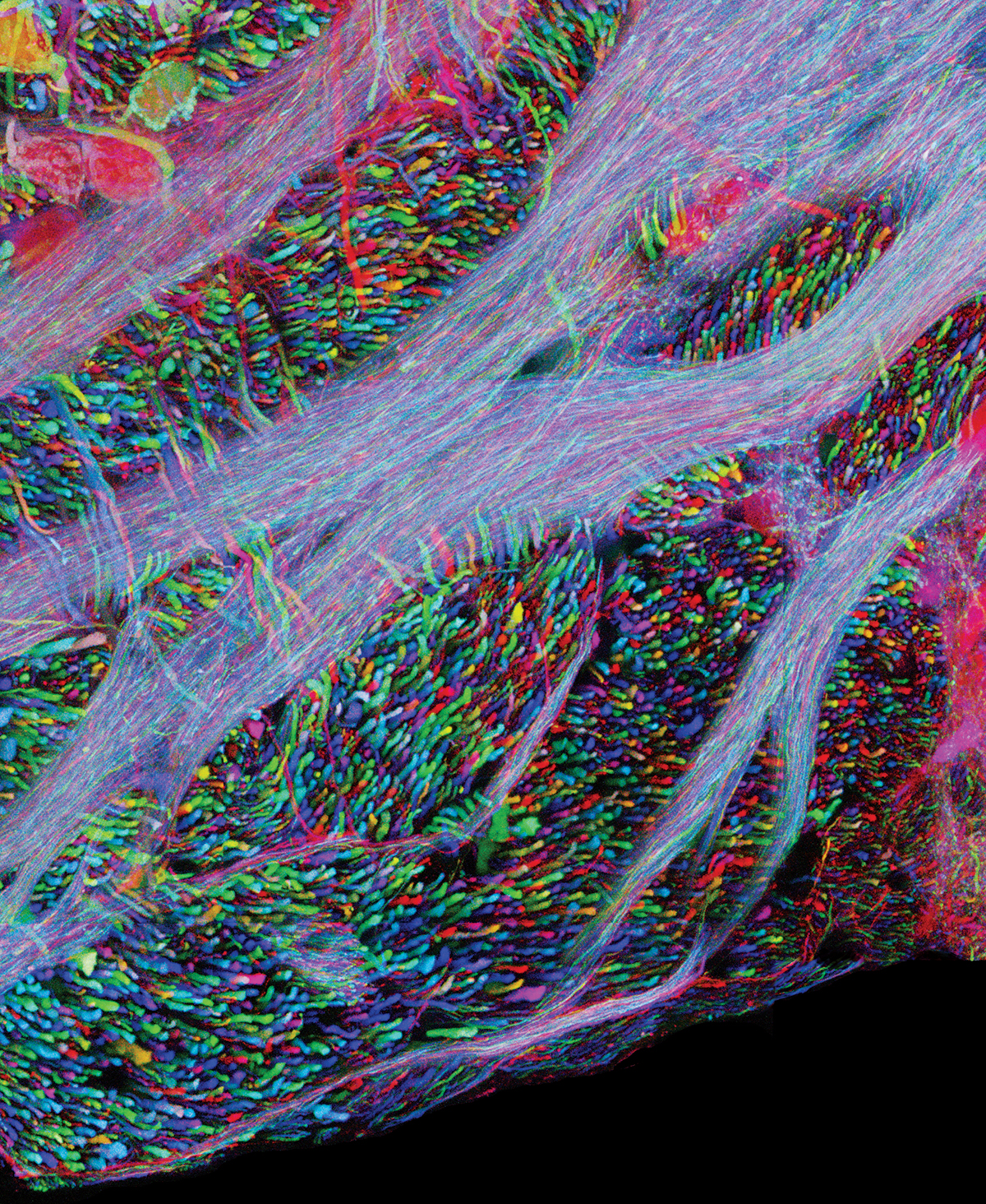
image courtesy of Cai, Lichtman, and Sanes, Harvard University
Axons in the auditory part of the mouse brainstem, imaged using the Brainbow technique.


